
On this page
ABOUT STONY CORAL TISSUE LOSS DISEASE
ABOUT WHITE PLAGUE DISEASE
WPD (White Plague Disease) & SCTLD (Stony Coral Tissue Loss Disease) are macroscopically similar
Because SCTLD and WPD are macroscopically similar and specific causative agents have not been identified for either disease, it is not a simple matter to confirm or not the presence of SCTLD in Barbados in early 2023. To distinguish between the two diseases we have to rely on information on patterns of spread of the diseases, and analyses of tissue samples combined with ongoing research on the nature of the associated microbial communitiese. e, g., see
– Similarities and Differences Between Two Deadly Caribbean Coral Diseases: White Plague and Stony Coral Tissue Loss Disease, Aldo Cróquer et al. 2021 in Frontiers in Marine Science
– Experimental transmission of Stony Coral Tissue Loss Disease results in differential microbial responses within coral mucus and tissue, Naomi Huntley et al.m 2022 in Nature ISME.
ABOUT STONY CORAL TISSUE LOSS DISEASE
Impacts of stony coral tissue loss disease on the persistence of Caribbean cleaner gobies
Budd et al., 2024 in Front. Mar. Sci., 03 April 2024 “Stony coral tissue loss disease (SCTLD) is the most recent of many diseases documented to impact Caribbean stony corals. SCTLD is known to impact over 20 species of reef-building corals and can cause complete colony mortality of large corals in only one month. Among the coral species impacted are those occupied as cleaning stations by Caribbean cleaner gobies. This study examined the persistence of these gobies on living coral cleaning stations where SCTLD was most recently or not yet affected (emergent), recently established (epidemic), and well-established (endemic)…This suggests that gobies are remaining at the site but may be abandoning live coral cleaning stations as the individual colonies are affected by SCTLD. Given the benefit cleaner gobies have on local coral reef fishes, changes in cleaning activity associated with coral disease have the potential to negatively impact Caribbean reef fish communities.”
Stony Coral Tissue Loss Disease in Florida Is Associated With Disruption of Host–Zooxanthellae Physiology
Landsberg et al., 2020 in Frontiers in Marine Science “… Collectively, pathology indicates that SCTLD is the result of a disruption of host–symbiont physiology with lesions originating in the BBW [basal body wall] leading to detachment and sloughing of tissues from the skeleton.”
A meta-analysis of the stony coral tissue loss disease microbiome finds key bacteria in unaffected and lesion tissue in diseased colonies
Stephanie M. Rosales et al., 2023. in ISME Communications. The introduction provides a concise review of what Is known about causes, spread of SCTLD
Threat of Stony Coral Tissue Loss Disease to Barbados
Page on website of Coastal Zone Management Unit (Barbados; accessed Jan 14, 2022).
“In the summer of 2014, corals off the coast of Florida were presenting with white blotches or bands associated with rapid tissue loss, leaving bare coral skeleton behind and killing corals within days to weeks. We at the CZMU have continued to monitor the progression of this disease, now known as the Stony Coral Tissue Loss Disease (SCTLD), as well as the emerging science surrounding the issue. Unfortunately it has reached our neighbours in St. Lucia and the possibility of it making its way to Barbados is slowly increasing. As we work on a plan of action should the disease reach our shores, we ask for your assistance in surveillance and reporting of any suspected cases of SCTLD. The posters below outline decontamination procedures as well as the basic characteristics of the disease.” (Read more)
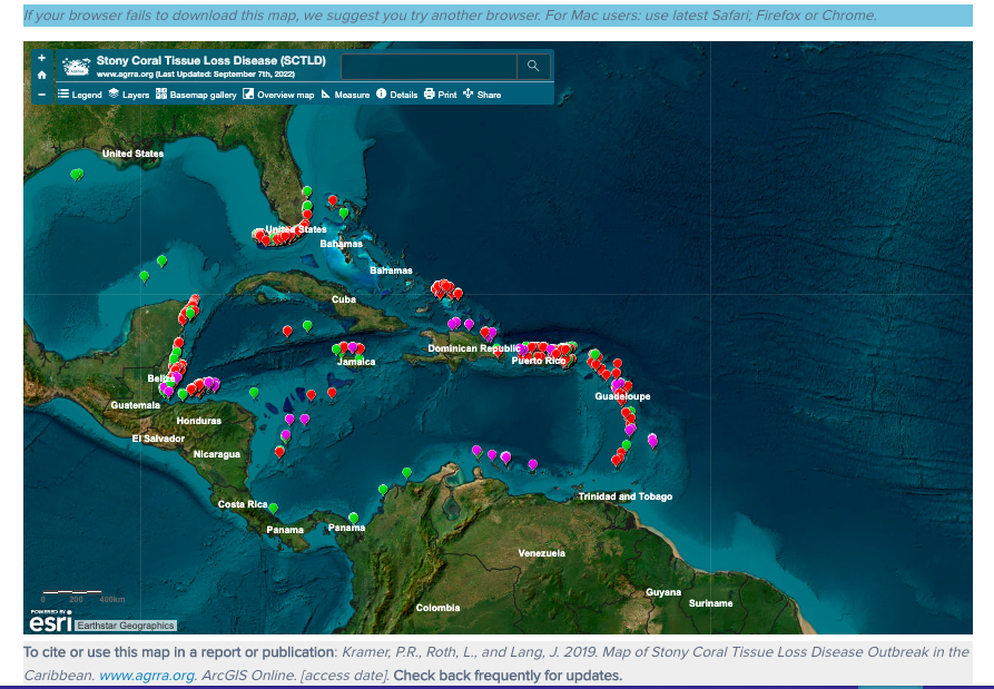
Screen capture of the interactive map
AGRRA: Stony coral tissue loss disease outbreak
AGRRA is the acronym for “Atlantic and Gulf Rapid Reef Assessment” Comprehensive. Includes interactive map for monitoring the disease.
AGRRA: Case Definition: Stony Coral Tissue Loss Disease (SCTLD)
Document listing species by susceptibility; photos etc. About Sidereastera siderea, it notes: S. siderea may show disease signs before highly susceptible species, during outbreaks, and after the outbreak has progressed through a reef system. The presentation of disease may be similar to SCTLD in some but not all cases, and the epidemiology, e.g., the patterns of lesion spread within and among colonies and duration of tissue loss, does not always match those of other species. This raises some uncertainty about inclusion of S. siderea in this case definition.
——————–
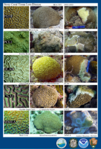
AGRRA Field Guide to Stony Coral Tissue Losses Disease
 – Card1 Front Two healthy coral pics and one diseased pic of: P. strigosa, P. clivosa. D labyrinthiformis, C. natans, M.meandrites.
– Card1 Back More diseased pics of P. strigosa, P. clivosa. D labyrinthiformis, C. natans, M.meandrites.
Card2 Front Two healthy coral pics and one diseased pic of Dichocoenia stokesii, Orbicella annularis, Orbicella faveolata, Montastrea cavernosa, Siderastera siderea
Card 2 Back: More diseased pics of of Dichocoenia stokesii, Orbicella annularis, Orbicella faveolata, Montastrea cavernosa, Siderastera siderea
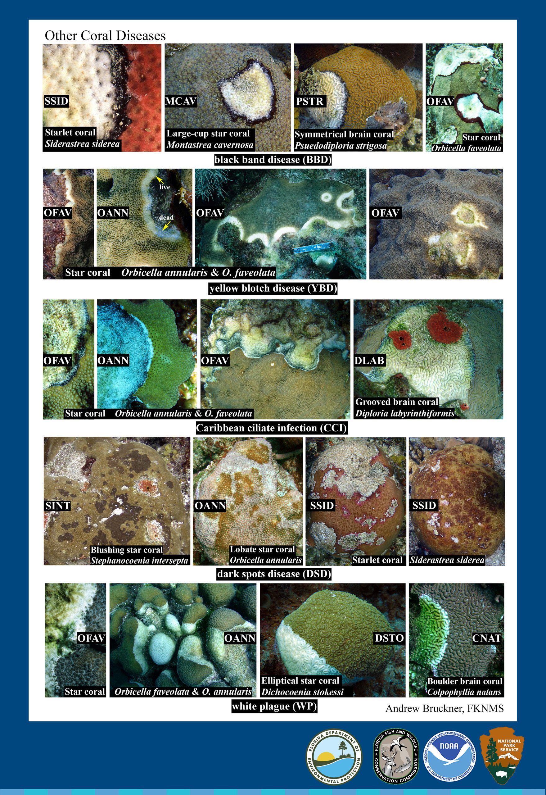
Card 3 Front: Other Coral Diseases
– Black Band Disease on S. siderea, M. cavernosa, P. strigosa, O. faveolata
– Yellow Blotch Disease on O. annularis & O. faveolata
– Caribbean Ciliate Infection on O. annularis & O. faveolata, & D. labyrinthisformis
– Dark Spots Disease on S. intersepta, O. annularis, S. siderea
– White Plague on O. faveolata & O. annualaris, D. stokessi, & C. natans
Card3 Back Other Conditions Affecting Corals– stages of coral bleaching, growth anomalies, bleaching & black band diseases, overgrwoth by sponges, other organisms, stoplight parrot fish predation, dameselfish ridge mortality, snail predation,
———————
– AGRRA: How do you recognize and describe Stony Coral Tissue Loss Disease (SCTLD) lesions?
Andy Bruckner, 2018. PDF of presentation, many photos of different diseases, grazing of corals etc.
———
Some of the highly susceptible species. Screen capture from AGRRA
——–
From AGRRA: Case Definition: Stony Coral Tissue Loss Disease (SCTLD) (2018)
Left: Highly Susceptible Species Early onset (the species first affected during an outbreak), rapid progression, and total mortality ranging from one week for smaller colonies to complete mortality over 1-2 months for larger colonies. Typically, M. meandrites and D. stokesii are the first to become affected at a site, followed by C. natans, and then the others show disease signs shortly thereafter.
Right: Intermediately Susceptible Species: Onset of tissue loss typically occurs about a month after onset in highly susceptible species, but lower numbers may also show disease signs before or as those species are affected. Smaller colonies die out over months, and larger colonies may show new lesions continuing with possible mortality occurring over years.
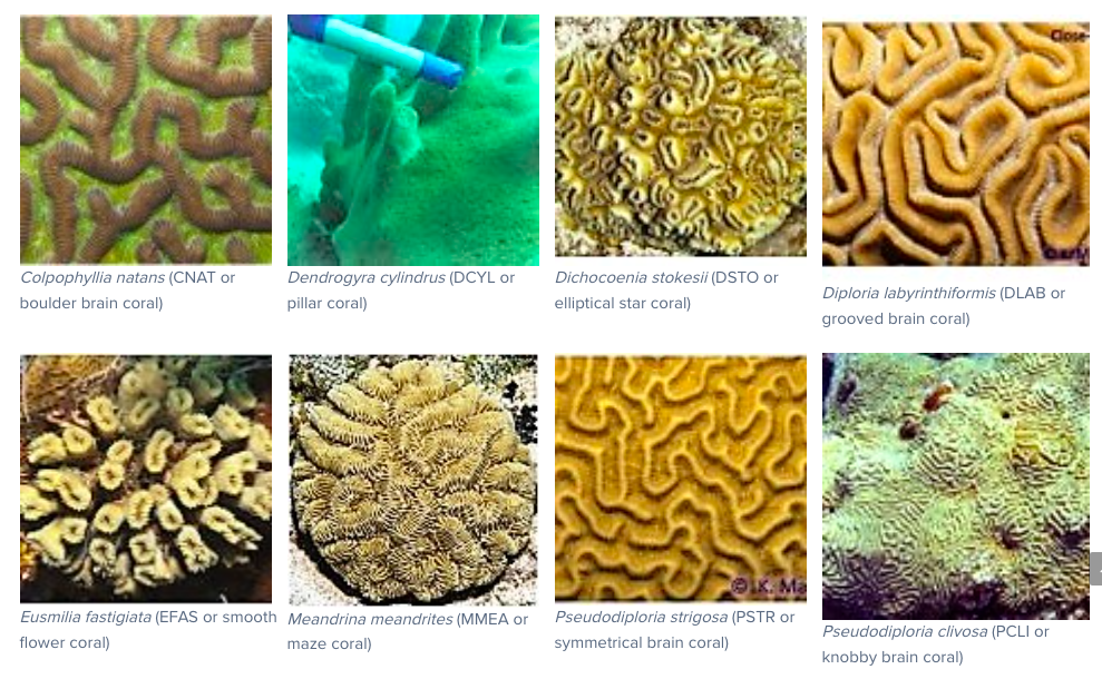
| Highly Susceptible Species Colpophyllia natans (boulder brain coral) Dendrogyra cylindrus (pillar coral)* Dichocoenia stokesii (elliptical star coral) Diploria labyrinthiformis (grooved brain coral) Eusmilia fastigiata (smooth flower coral) Meandrina meandrites (maze coral) Pseudodiploria strigosa (symmetrical brain coral) Pseudodiploria clivosa (knobby brain coral) |
Intermediately Susceptible Species Orbicella annularis (lobed star coral)* Orbicella faveolata (mountainous star coral)* Orbicella franksi (boulder star coral)* Montastraea cavernosa (large-cup star coral) Solenastrea bournoni (smooth star coral) Stephanocoenia intersepta (blushing star coral) Siderastrea siderea (starlet coral)** |
Left: Presumed Susceptible & Right: Low Susceptible Species
| Presumed Susceptible but insufficient data to categorize onset. Agaricia agaricites (lettuce coral) Agaricia spp. (plate/saucer corals) Mycetophyllia spp. (cactus coral) Madracis arenterna (pencil coral) Favia fragum (golfball coral) Helioseris cucullata (sunray lettuce coral) Mussa angulosa (spiny flower coral) Scolymia spp. (disc coral) Isophyllia spp. (sinuous cactus coral; rough star coral) |
Low Susceptible Species: During outbreaks, the following corals are rarely or not affected. From Case Definition: Stony Coral Tissue Loss Disease (SCTLD) (cited above): Porites astreoides (mustard hill coral) P. porites (finger coral) P. divaricata (thin finger coral) P. furcata (branched finger coral Acropora palmata (elkhorn coral)* A. cervicornis (staghorn coral)* Oculina spp. (bush corals) Cladocora arbuscula (tube coral) |
*Endangered Species Act (ESA) listed species
——–
NOAA releases Stony Coral Tissue Loss Disease response plan
US: NOAA Oct 5, 2022
Stony Coral Tissue Loss Disease, first identified in 2014, has harmed more than 22 species of stony corals in Florida, the U.S. Virgin Islands and Puerto Rico, and continues to spread across the Caribbean. Cases have been confirmed in at least 20 countries and territories.
The outbreak is unique in its rapid progression, high death rates, large geographic range, extended duration and the number of coral species affected. Once infected, coral colonies typically die within weeks to months.
Today, NOAA is releasing a plan to guide future actions to treat and prevent the spread of a disease affecting coral reefs in Florida and the U.S. Caribbean. The plan also includes actions to prevent the spread of the disease to the Indo-Pacific.
Details at Stony Coral Tissue Loss Disease (NOAA Coral Reef Information System)
———
Stony coral tissue loss disease decimated Caribbean coral populations and reshaped reef functionality
Lorenzo Alvarez-Filip et al., 2022 in Nature. “…The outbreak was first reported in Florida in 2014 and reached the northern Mesoamerican Reef by summer 2018, where it spread across the ~450-km reef system in only a few months. Rapid spread was generalized across all sites and mortality rates ranged from 94% to <10% among the 21 afflicted coral species. Most species of the family Meandrinadae (maze corals) and subfamily Faviinae (brain corals) sustained losses >50%…This emergent disease is likely to become the most lethal disturbance ever recorded in the Caribbean, and it will likely result in the onset of a new functional regime where key reef-building and complex branching acroporids, an apparently unaffected genus that underwent severe population declines decades ago and retained low population levels, will once again become conspicuous structural features in reef systems with yet even lower levels of physical functionality.
From Methods : We focused on exploring the prevalence and geographical and temporal trends of the most ‘highly susceptible species’, which were species with more than 10% disease prevalence (considering diseased and recently deceased colonies). These species are:
ABOUT WHITE PLAGUE DISEASE
White Plague Disease (Wikipedia)
“White plague is a suite of coral diseases of which three types have been identified, initially in the Florida Keys. They are infectious diseases but it has proved difficult to identify the pathogens involved. White plague type II may be caused by the gram negative bacterium Aurantimonas coralicida in the order Hyphomicrobiales but other bacteria have also been associated with diseased corals and viruses may also be implicated…In 1977, a disease of scleractinian corals appeared on reefs off the Florida Keys in the United States and was termed white plague…” Good overview
Aldo Cróquer et al. 2021. Similarities and differences between two deadly Caribbean coral diseases: White plague and stony coral tissue loss disease. In Frontiers in Marine Science. Full article is available.
Coral Disease & Coral Bleaching
Factsheet from Florida Dept of Environmental Protection, Aug 2017. “White plague is a bacterial infection that is often confused with coral bleaching. Recent studies suggest
that there are many variations of the disease. In all cases, corals exhibit loss of tissue, leaving the bare, white skeleton exposed. Type I White Plague, also commonly known as white band disease, occurs more frequently on branching corals, starting at the base of the branches and moving towards the tips at a rate of a few millimeters per day Type II White Plague moves
similarly to Type I but typically in the opposite direction, and is more common in boulder or massive coral species.
 Coral disease ravages reef-building corals throughout Southeast Florida
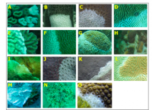
William F. Precht et al., 2018 on insideecology.com. Panel shows disease on 15 coral susceptible coral species. “Species are listed in decreasing order of prevalence/mortality where (A) was most heavily impacted and (O) was the least impacted. Species include: (A) Eusmilia fastigiata, (B) Dendrogyra cylindrus, (C) Dichocoenia stokesi, (D) Meandrina meandrites, (E) Colpophyllia natans, (F) Pseudodiploria strigosa, (G) Diploria labyrinthiformis, (H) Orbicella annularis, (I) Solenastrea bournoni, (J) Montastraea cavernosa, (K) Orbicella faveolata, (L) Pseudodiploria clivosa, (M) Mycetophyllia aliciae, (N) Oculina diffusa, and (O) Orbicella franksi.”
White Plague is the New Black Plague
By: Julia Paoli 2014 On Nature Scitable “White plague is debilitating to coral reefs. It causes rapid tissue loss in corals, which leaves their white skeletons exposed to the elements. Multiple species of coral are affected by white plague. Ultimately, the disease can cause partial or complete colony mortality in corals. Soffer says that white plague is not difficult to spot; “You have living, healthy tissue, and then immediately below that you have a straight band of white on the bottom of the coral.” The white band can spread quickly to the rest of the colony.
“Scientists have identified three types of white plague that differ only in the rate they spread. The first kind identified in 1977 is known as Type I and destroys roughly one tenth of an inch (3 millimeters) of coral tissue per day. Type II is more lethal, with a mortality rate of .8 of an inch (2 centimeters) per day. At this rate Type II can annihilate a small colony in one to two days. The most recently discovered kind is Type III, which was first found in 2000. Type III kills coral tissue at a rate greater than .8 of an inch a day and mainly targets large reef-building corals.” Article goes on to comment on research on possible causative agents