
Drafting Apr 18, 2023…
Long Title: Progression of coral diseases on Vauxhall Reef & Environs Jan 11 to Apr 6, 2023
January 11, 2023: Conditions were perfect for snorkelling at the Vauxhall Reef and I looked out for SCTLD-infected corals on a route approx. perpendicular from shore that took me across the Reef Flat, Diploria-Palythoa, Reef Crest, Seaward Slope and Reef Front zones (Lewis 1960), to ‘The Wreck”. I saw only one obviously infected coral, that in the outer Reef Crest zone, until I reached The Wreck where there were many, and at least some of them appear to fit the description for Stony Coral Tissue Loss Disease.
Jan 12-21, 2023: I have criss crossed the Vauxhall Reef out to & including the Reef Front several times, and swum along the seaward edges of the next 2 fringing reefs to the north, finding about 30 “Obviously Infected Corals” in total but every time I look, I find more. Currently, most of the infected corals (i.e., with symptoms characteristic of SCTLD) appear to be Pseudodiploria strigosa, Colpophyllia natans, Diploria labyrinthiniformis, Meandrites meandrina: possibly some specimens are infected.”
From Aldo Cróquer et al. (2021), bolding inserted:
“The unique feature that appears to be exclusive to SCTLD is the multifocal, coalescent lesions that spread across the affected colonies (Aeby et al., 2019) and a front of sloughing tissue (Supplementary Material), whereas WPD starts from the colony base and progresses upward in a concentric ring or elongated band (Weil et al., 2002; Sutherland et al., 2004; Weil et al., 2006; Bruckner, 2016b; Richardson, 2016; Figures 3A–F). Nevertheless, multifocal lesions were also described as an “occasional” condition for WPD-II (Richardson et al., 1998b), and the condition was observed in different geographic localities in the 2000s (Weil and Croquer pers. Observations; Figures 4G–J). Thus, the distinction between macroscopic signs of SCTLD and WPD is challenging.”
Most of the OICs (Obviously Infected Corals observed over the interval Jan 11-21 seem to fit the description of WPD, rather than SCTLD. A few had multifocal lesions not starting from the base, but “a front of sloughing tissue” was not observed on any OIC (I looked for it). View photos under AGRRA Rep 1 (Fore Reef, Jan 21, 2023); AGRRA Rep 2 (Fore Reef, Jan 11, 2023); AGRRA Rep 3 (Wreck on Fore Reef,Jan 14, 2023).
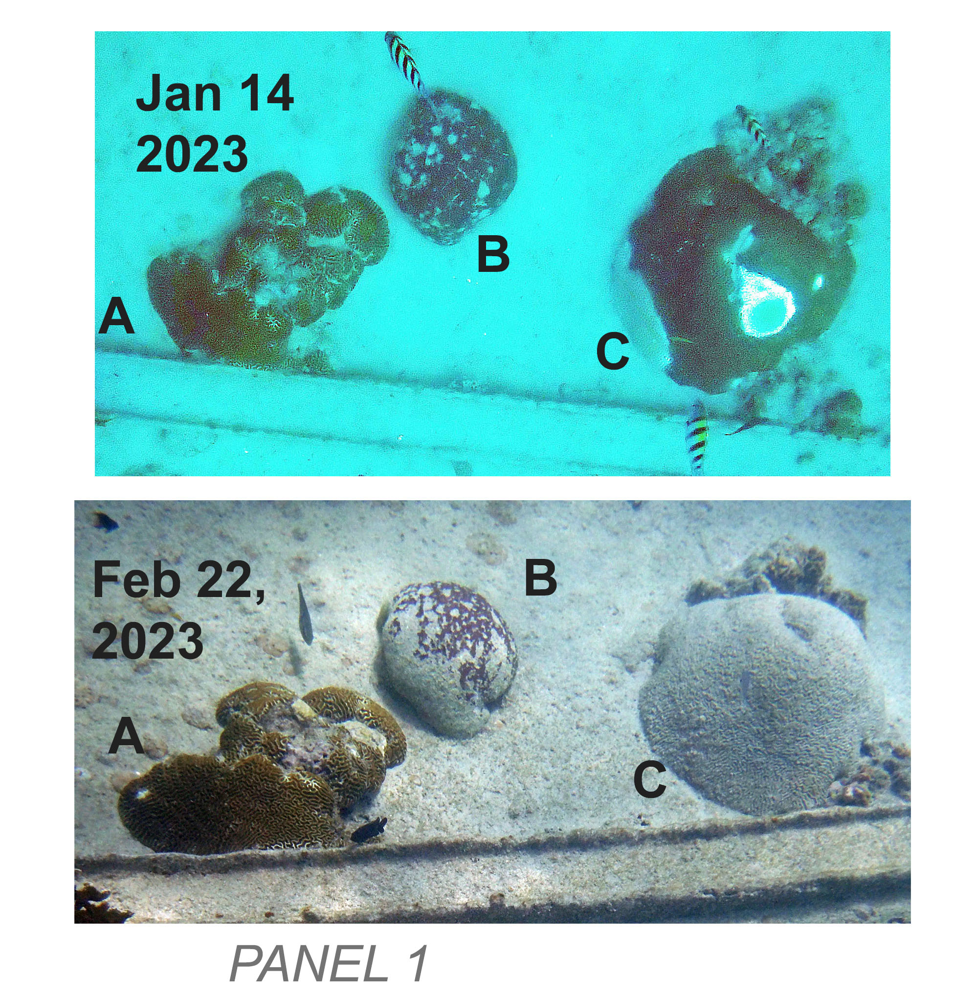
When I observed corals on The Wreck early on, there were some that were completely white/covered with fine sediment. At the time, I wasn’t sure whether those might have been corals that died some time ago from other causes, e.g. coral bleaching. In AGRRA Rep 15 (Feb 20, 2023), AGRRA Rep 17 (Feb 22, 2023), and AGRRA Rep 18 (Feb 24, 2023), I photographed corals on the wreck that had been photographed on Jan 14 or 15th; these illustrated rapid progression of the disease including a P. strigosa specimen that went from circa 25% disease cover on Jan 14 to completely dead and covered by fine white sediment on Feb 22. The earlier photo illustrates multifocal infection on this specimen. By April, there were many individual corals that had earlier been uninfected or partially infected that were then completely dead and covered with fine white sediment (details/photos not yet posted). So these observation suggest that the completely dead corals covered with fine white sediment observed on Jan 14 & 15 (most appeared to be P. strigosa) were likely infected at least one month earlier.
‘Still workin on it